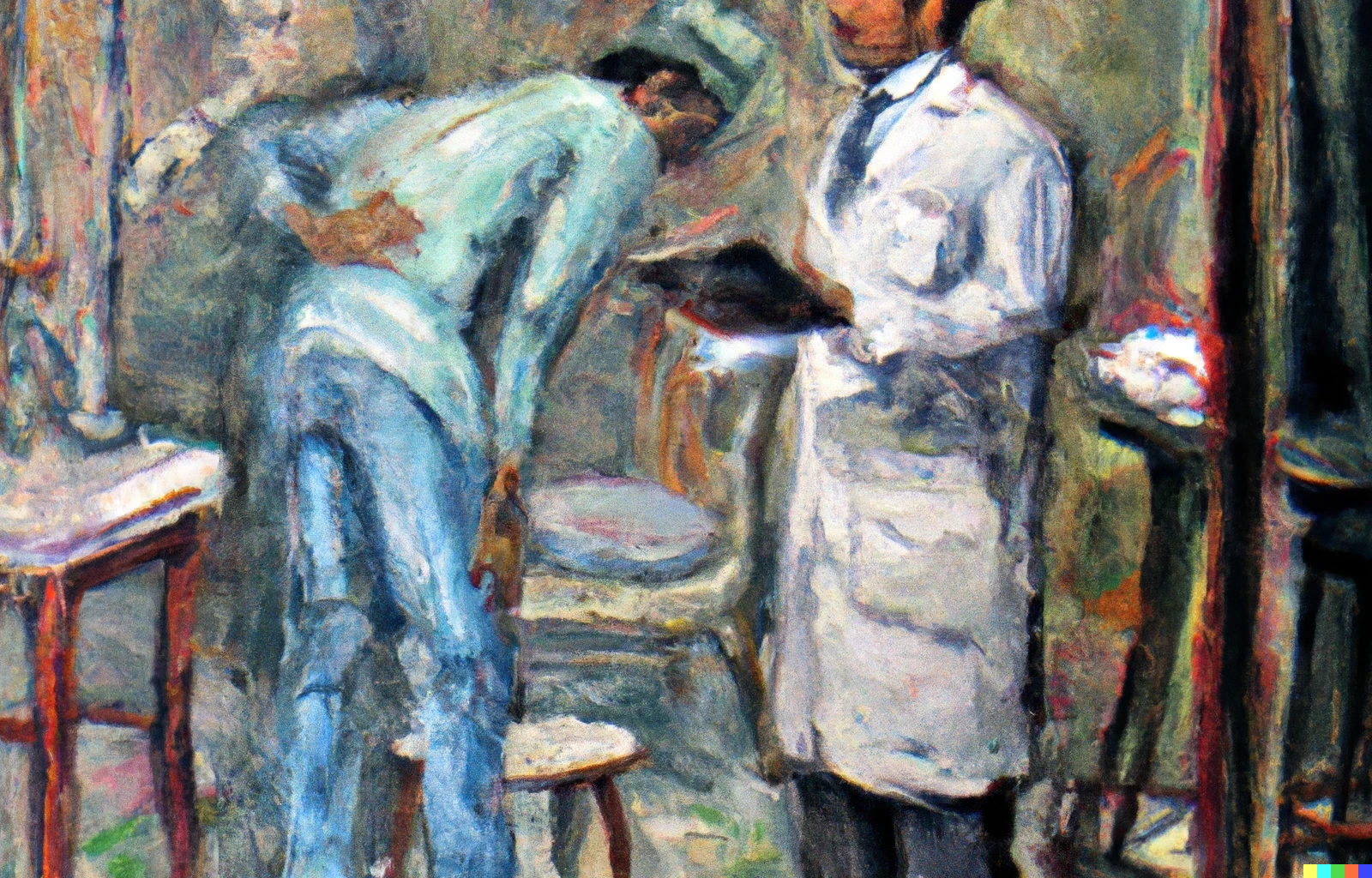
Lessons Learnt from The Translumbar Aortogram
Any image guided biopsy can cause two major complications. The first is bleeding, which can happen from within a vascular organ (liver, spleen, kidney) or when you injure or traverse a vessel (lung, liver, abdomen, head & neck, mediastinum). The second is a pneumothorax.
Many radiologists balk at the idea of biopsying lesions close to the aorta or its branches, fearing that if they lose control, the needle may puncture the aorta or another artery and cause catastrophic bleeding. I always tell the younger radiologists and residents that unless you land up biopsying the wall of the vessel itself, the simple puncture of a vessel is unlikely to ever cause catastrophic bleeding, assuming that the coagulation parameters are normal.
In any case, transvascular, transaortic biopsies and injections have been described for some years now. So why would you worry about a needle inadvertently just entering a vessel? And…if you have ever seen or done a translumbar aortogram (TLA), you will never again worry about puncturing the aorta.
I was a resident from 1988 to 1991 and one of the first procedures that I saw and eventually performed in my 1st and 2nd years was the TLA…until we mastered the Seldinger technique in the latter half of my second year and started doing transfemoral aortograms.
You have to picture this in your head.
Two or three of us are in the neuroradiology suite, using a MIMER III neuro X-ray machine with a manual cassette changer, without the fluoroscopy attachment, which stopped working a few years ago. The patient is prone. One of us, let’s say it is me, palpates the spinous processes and approximates the L3/L4 level. I pick a space between two transverse processes, a few centimeters from the midline and blindly insert an 18G metal autoclaved needle in the general direction of the aorta, until I feel a give-way that tells me I have entered the aorta. Another way is to angle the needle in the direction of the vertebral body until you touch bone, then realign the needle a little more laterally and puncture the aorta. I then remove the stylet. If I am in the aorta, there will be a fountain of blood spurting under pressure from the needle (a standard joke was that the discoloration on the ceiling of the X-ray room was because of blood from some patients that had reached that height), in which case I put the stylet pack. If there is no spurt, chances are I have gone beyond the anterior wall, so I withdraw a little and then hope to get a spurt.
When the needle is in the aortic lumen, you can see it moving wildly with each aortic pulsation and we need to move fast…our hearts are also beating a little wildly.
Now we have to inject ionic iodinated contrast (Conray 420) using a 50 cc glass syringe. If an O2 cylinder is ready, then we use a contraption to push the plunger with the force of the gas. Most of the times, this device is under repairs and so the strongest person in the room attaches the syringe to the needle with some tubing and gets ready to inject the contrast. If you are a resident or a consultant who has passed out in the last 10 years, I understand that you may have never felt contrast medium with your fingers, leave alone injected contrast into patients, which is nowadays almost always done by nurses or trained non-doctor personnel. But if you have felt iodinated non-ionic contrast, then think of something 5-10 times more viscous that has to be injected with force…and you will understand the strength needed to do this as fast a possible, with a glass syringe and a metal needle. You also have just one shot to get everything right for that one aortogram image.
Before the injection, we make one final check of the cassettes and reconfirm that they are loaded with films. We have to make sure that the X-ray generator has been switched on, that someone is ready behind the lead curtain to take a shoot when we shout “shoot” and that another technician or often just one of us radiology residents is ready with the cassettes, which we have to manually change after each shoot, as fast as possible, so that we can get at least a couple of films with contrast in the aorta.
With a deep breath, I ask the now irritated technician to recheck everything. I tell the patient not to move under any circumstances, and I pray there is no instant contrast reaction. The injection starts. At around 30 seconds or when 50-60% of the contrast has been injected, we yell in concert, “shoot”. The technician shoots and immediately, my colleague pulls the cassette changer, removes the cassette and puts a new one and pushes the changer in and yells, “shoot” and the tech shoots again. Chances are the injection is just nearing completion by then. Just for safety, another shoot is taken.
With the needle still in, someone rushes with the cassettes to the dark-room and starts developing the films to check whether there is an adequate image of the aorta. If yes, they shout from inside that it’s all good and I remove the needle and turn the patient over. If they yell no, then often times, the entire procedure is repeated once again.
All this when everything works in sync. If we do get a successful aortogram image, we look up and thank whoever it is, who is looking out for us…because half the times, the X-ray tube will not shoot or the X-ray generator will trip or the piston will get stuck in the glass syringe or the syringe will break or the cassette tray will get stuck or somehow it will just take a very long time to change the cassette, or we will mistime the shoot or no one will shout “shoot” because each one thinks the other is going to, or the electricity will suddenly go off just at the time of injection or the patient will move violently or the cassettes will be found empty, because someone forgot to put films inside.
If we did get one or two films showing the aorta well, we would march triumphantly with “wet” films on hangers down to the reporting area, show off to all our colleagues there and then figure out whether the aorta was normal or abnormal and then write out a report by hand after checking with our seniors...and then communicate the findings to the referring physician or surgeon.
At the end of all this, we would then troop to the canteen, have a few cups of cutting chai, feel good about ourselves and happy that we had been successful…and then we would call it a day.
We never knew how much the patients bled when we withdrew the needle because we had no ultrasound or CT scan to check…but I do remember that we did not lose any patient to shock or bleeding. Our referring doctors also had our backs and used to encourage us to do these procedures and give us the confidence that if anything went wrong, they would take care of the patient.
If you have done all this including direct carotid puncture angiograms for head injuries, which I will describe another time, you never should have to worry about putting a needle into an artery by mistake or on purpose. By extension, all of you who put needles inside body parts should never refuse biopsies of nodes or masses close to the aorta or any of the great vessels, just for fear of puncturing the vessel.
More Reading:
These two articles describe the MIMER and MIMER 3, just in case you would like to know how it was 50-75 years ago. You can download and read them.
This Siemens article also has an image showing a patient in the MIMER 3 machine.
Acknowledgments:
Thanks to Dr. G. R. Jankharia for sharing his memories of the TLA in Nair Hospital, Dr. Ravi Ramakantan for his edits and inputs, Dr. Anand Parihar and Dr. Gurmit Singh for clarifying some technical issues and Dr. Milind Gune, my partner-in-crime at Sion Hospital (LTMMC and LTMGH) for confirming these 35-years old memories and remembering some more.
Bhavin's Writings Newsletter
Join the newsletter to receive the latest updates in your inbox.


